
Imaging is a commonly used tool in professional sport for Sports Injuries. It is also accessible to the general public for Sports and Musculoskeletal injuries.
Imaging is used to get more information on an injury after an initial clinical assessment has been done by your Sports Physio or Doctor.
Common imaging options include X-Ray, MRI (Magnetic Resonance Imaging) and CT (Computed Tomography) scans.
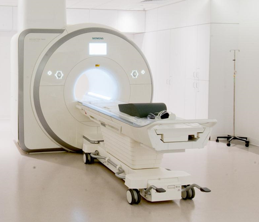
MRI Machine
What Is Imaging?
Medical imaging is most commonly done in private radiology clinics or within hospitals.
There are many different types of imaging that allow us to see our musculoskeletal anatomy that assist in an injury diagnosis.
This can be useful in many types of injuries, especially if injury diagnosis is unclear after standard clinical assessment.
Why Is Imaging Used?
Imaging can be used to give further information about an injury, or to confirm or exclude a diagnosis.
Imaging for sports injuries can give important information that directs the type of management and treatment of an injury. In all cases imaging results need to be interpreted by an experienced Sports Doctor or Sports Physiotherapist and used in conjunction with your clinical presentation and symptoms.
Types of Imaging
The most common types of imaging used for sports are;
X-Ray – This is the easiest, most affordable and most commonly used. X-rays give good information on bone injuries such as fractures, and also some information about joints and swelling eg. Knee Osteoarthritis.
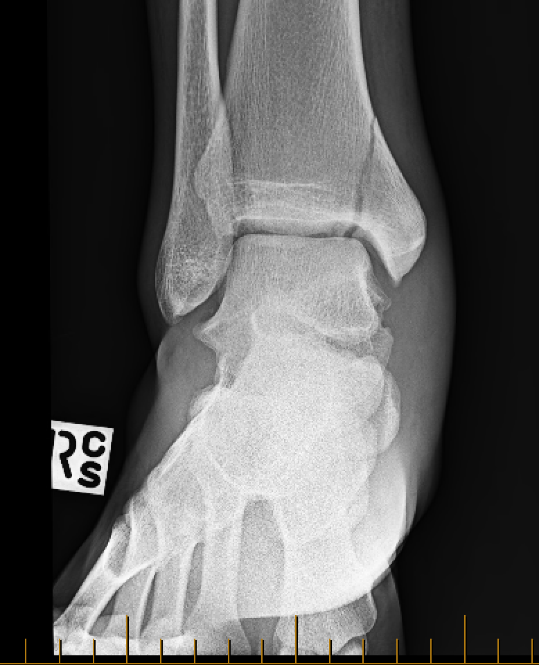
Ankle fracture on X-Ray
MRI – Gives great information about soft tissue injuries such as muscle strains and ligament sprains. These scans can be very useful in assisting diagnosis and prognosis of injuries.
They also provide further insight into hard tissue such as cartilage and bone.

Knee MRI - Green arrows pointing to Meniscus cartilage tear
CT – Used for more detailed information on bone. Commonly used when an X-Ray provides a negative result but a fracture or bony injury is still suspected.
What Injuries Are Scans Useful For?
Scans can provide crucial information for both acute and longer-term chronic injuries. Some examples of these include hamstring and calf muscle strains, knee and ankle ligament sprains, eg. ACL/MCL, and for acute fractures.
Scans give information about grading of these injuries, which is important to decide how each injury is managed, including whether a rehab period is appropriate or referral to a specialist or surgery is needed.

MRI - Green line and surrounding white fluid indicating Hamstring Strain
Once you have had a scan it is important to consult with your Sports Physio or Sports Doctor to get an accurate assessment and diagnosis of your injury.